You are currently browsing the archives for the cool tools category.

Archive for the ‘cool tools’ Category

A Revolution in Mapping Complex Genetic Traits

 :: Posted by American Biotechnologist on 09-18-2014

Life is rarely simple. From crop yields to disease risks, the biological characteristics people care most about are usually those considered “complex traits.” Just as for height—the textbook example of a complex trait—attributes like risk for a particular human disease are shaped by multiple genetic and environmental influences, making it challenging to find the genes involved. To track down such genes, geneticists typically mate two individuals that differ in key ways—for example, a large mouse and a small mouse—and then study their descendents, looking for genes that tend to be inherited with the trait value of interest. But this method only implicates a broad genomic region, and the identities of the crucial gene/s often remain a mystery.

Now, geneticists are embracing a powerful approach that pinpoints more precise areas of the genome by founding the breeding population with multiple, genetically diverse parents. To encourage innovations in this rapidly developing field, the Genetics Society of America journals GENETICS and G3: Genes|Genomes|Genetics today published the first articles in an ongoing special collection on mapping complex trait genes in multiparental populations.

The 18 articles describe methods and applications in a wide range of organisms, including mice, fruit flies, and maize. Among the advances reported are the creation of a multiparental population of wheat, methods for use with the Diversity Outbred and Collaborative Cross mouse populations, and the identification of nicotine resistance genes in fruit flies. The power of the approach for disease genetics is highlighted in an article describing how a multiparental rat population was used to find a human gene variant that affects insulin levels.

“These collections of multiparental strains are extremely powerful and greatly accelerate discovery. For example, in one of the articles, researchers report using a multiparental population to rapidly identify fruit fly genome regions associated with the toxicity of chemotherapy drugs. The authors could then examine these regions to find several candidate causative genes,” said Dirk-Jan de Koning, Professor at the Swedish University of Agricultural Sciences, Deputy Editor-in-Chief, Complex Traits, at G3, and an editor of the new collection. “Using standard two-parent crosses, they would have been stuck with unmanageably large regions each containing hundreds or even thousands of candidate genes.”

Because the field is so new, geneticists are still developing the best methods for creating and analyzing multiparental populations. “This collection will move the field forward by stimulating discussion between different disciplines and research communities,” said Lauren McIntyre, Professor at the University of Florida, and an editor of the collection. “To help foster this ongoing exchange, the collection will continue to publish new articles, and all associated data will be freely available.”

In an editorial, McIntyre and de Koning describe how the idea for the multiparental populations collection was born and how scientific society journals like GENETICS and G3 can advance new research fields.

Thanks to Genetics Society of America for contributing this story.

A Revolution in Pipetting

 :: Posted by American Biotechnologist on 08-05-2014

A team of Whitehead Institute researchers is bringing new levels of efficiency and accuracy to one of the most essential albeit tedious tasks of bench science: pipetting. And, in an effort to aid the scientific community at large, the group has established an open source system that enables anyone to benefit from this development free of charge.

Dubbed “iPipet,” the system converts an iPad or any tablet computer into a “smart bench” that guides the execution of complex pipetting protocols. iPipet users can also share their pipetting designs with each other, distributing expertise across the research community. The system, created by researchers in the lab of Whitehead Fellow Yaniv Erlich, is described in detail in a letter appearing this week in the journal Nature Methods.

Erlich says that today’s experiments frequently rely on high-throughput methods that combine large numbers of samples with large scale, complex pipetting designs. Pipetting errors can lead to experimental failure. Although liquid-handling robots would seem to be a logical choice for such work, they are also extremely expensive, difficult to program, and require trained personnel. Moreover, they can be plagued by technical snafus, ranging from bent or clogged tips to an inability to capture liquids lying close to the bottoms of individual wells.

“We needed an alternative to costly robots that would allow us to execute complex pipetting protocols,” says Erlich. “This is especially important when working with human samples that are often in limited supply.”

iPipet illuminates individual wells of standard 96- or 384-well plates placed on top of a tablet screen, guiding users through the transfer of samples or reagents from source to destination plates according to specific designs. Users create their own protocols in Microsoft Excel files in comma-separated format and upload them to the iPipet website, which generates a downloadable link for execution on a tablet computer. Included on the iPipet site are a variety demos and an instructional video.

So, how well does iPipet work? Beautifully, according to members of the Erlich lab. In a test of the tool against a liquid-handling robot, iPipet enabled nearly 3,000 fixed-volume pipetting steps in approximately seven hours. After significant time spent on calibration, the robot accomplished only half that number of steps in the same allotted time. To date, one of the only challenges lab users have encountered is keeping well plates in a fixed position on the tablet screen. For that, Erlich’s team provides a solution: a 3D printed plastic adaptor that users can create with a file accessible via the iPipet website.

“The entire iPipet system is open source,” says Erlich. “We want to maximize the benefit for the community and allow them to further develop this new man-machine interface for biological experiments”

Thanks to Whitehead Institute for Biomedical Research for contributing this story.

A Revolution in Scientific Publication

 :: Posted by American Biotechnologist on 07-30-2014

Since we are talking about impact factors and Journal related stuff, (see When JIF Becomes a Dirty Word), I wanted to share with you a very cool concept that I saw recently in F1000 Research.

Aside from it’s move to the digital world, scientific publication, as we know it, has remained relatively constant for over four hundred years. Papers are written in a scientific method-based theme and broken down into bite size sections. Papers are very much there for scientists to communicate their findings with us and for the investigators to provide us with their personal interpretation of the data. While a sort of 2-way communication often happens via editorials and personal communication, the presentation of the data remains static and one dimensional. Results, which represent the heart of the researchar, often presented in tabular or pictorial format. Much of the effort and funding allocated to a research project can be distilled down to several figures and maximizing the communicative ability of these results is essential to successful publication. That is why the methodology used to publish a recent paper in the journal F1000 Research may, in fact, revolutionize the world of scientific publishing.

In the newly released article, German professor of neurogenetics, Bjorn Brembs, published a proof-of-concept figure allowing readers and reviewers to run the underlying code within the online article. Instead of presenting readers with a static figure that can only be interpreted by the author, Dr. Brembs submitted the figure’s underlying code to the journal, allowing readers and reviewers to render the figure in various formats giving them more control over interpretation of the original data.

According to Brembs, the ultimate goal is to set up all journal submissions in such a way that authors will no longer have to deal with figures. They will simply need to submit text with links to data and code, and the rest will be up to the reader.

The recent rise in retraction rates of scientific articles proves that attempts at reproducibility by other labs are crucial to cross-checking our understanding of science. With only one or two figures to choose from in the past, authors were incentivized to pick the view of the data that best demonstrated their conclusions. “The traditional method of publishing still used by most journals today means that as a referee or reader, the data cannot be reused nor can the analysis be checked to see if all agree with the reported conclusions”, said Brembs. “This slows down scientific discovery. We are pleased to be able to pioneer these two interactive figures with F1000Research, which will hopefully be the start of a big shift in the way journals treat their figures.”

Cell Isolation - The Two Worlds of Cell Separation

 :: Posted by American Biotechnologist on 06-30-2014

A Simple Way to Label Your Own Antibodies

 :: Posted by American Biotechnologist on 03-13-2014

Bio-Rad Laboratories announced the launch of its ReadiLink antibody labeling kits, one of the market’s simplest antibody conjugation solution for labeling microscale amounts (50–100 µg) of antibody. These kits are ideal for researchers interested in labeling their own antibodies for flow cytometry and cell sorting applications.

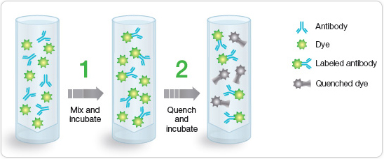
Using the ReadiLink antibody labeling kits, researchers can label their antibodies in two easy steps. The protocol takes only 70 minutes, and the labels are as bright and photostable as traditional dyes. Bio-Rad offers 10 different fluorophores whose excitation/emission wavelengths range from UV to infrared.

ReadiLink antibody labeling kits permit fluorophore conjugation of antibodies in two simple steps.

“The labeling kits will benefit a wide range of researchers,” said Mary Ferrero, Bio-Rad Product Manager in the Gene Expression Division, Life Sciences Group. “ReadiLink antibody labeling kits are ideal for researchers who are using a rare antibody, are interested in an antibody that is not commercially available with the appropriate fluorophore, or who want to label an antibody with a fluorophore that will fit into their multicolor flow cytometric experiment.”

Bio-Rad also offers additional reagents and consumables for each step of flow cytometry experiments, such as the ReadiDrop™ cell viability assays and primary antibodies.

For more information about Bio-Rad’s antibody labeling kits, please visit www.bio-rad.com/antibodylabeling.